
Introduction
Tissue sectioning accuracy and quality are critical in the fields of pathology and histology. This process, traditionally performed manually by skilled technicians, involves slicing thin sections of tissue samples for microscopic examination. With the advent of automated tissue sectioning, also known as automated microtomy, the field has witnessed a significant leap in precision, efficiency, and reproducibility. This article delves into the importance of automated tissue sectioning, its technological advancements, and its impact on medical research and diagnostics.
Definition
Slices (sections) of biological tissue are cut very thinly, usually for microscopic analysis. This procedure is known as tissue sectioning. Studying the internal structure, makeup, and cellular organisation of tissues is made possible by this method, which helps pathologists, biomedical researchers, and researchers in other domains including pathology and histology with diagnostic and investigative tasks.
The Need for Precision in Tissue Sectioning
Tissue sectioning is a critical step in the preparation of samples for histological analysis. The thin slices of tissue, typically ranging from 2 to 5 micrometers in thickness, must be uniform and free of artifacts to ensure accurate diagnosis and research outcomes. Manual sectioning, while effective, is prone to variability due to human factors such as fatigue, skill level, and consistency. Even the most experienced technicians can encounter challenges in maintaining the high standards required for clinical and research applications.
Advancements in Automated Tissue Sectioning
Automated tissue sectioning systems have emerged as a game-changer in histology laboratories. These systems leverage advanced technology to enhance the precision and efficiency of the sectioning process. Here are some key advancements:
Robotic Precision:
Automated microtomes are equipped with robotic arms and computerized controls that ensure consistent and precise cuts. These machines can be programmed to slice tissue samples at specific thicknesses, minimizing the variability associated with manual sectioning. The robotic precision not only improves the quality of the sections but also reduces the likelihood of human error.
Advanced Blade Technology:
The blades used in automated tissue sectioning systems are made from high-quality materials such as diamond or tungsten carbide. These blades are engineered to maintain sharpness over extended periods, ensuring clean and smooth cuts. Some systems also feature automatic blade-changing mechanisms, reducing downtime and enhancing workflow efficiency.
Automated Sample Loading and Unloading:
Automated tissue sectioning systems often include features for automated sample loading and unloading. This automation reduces the need for manual handling, minimizing the risk of contamination and damage to the samples. Additionally, it speeds up the overall sectioning process, allowing for higher throughput in busy laboratories.
Integration with Imaging Systems:
Modern automated microtomes can be integrated with digital imaging systems, enabling real-time visualization and quality control of tissue sections. This integration allows technicians to monitor the sectioning process and make adjustments as needed, ensuring that the sections meet the required standards for analysis.
Benefits of Automated Tissue Sectioning
The adoption of automated tissue sectioning offers numerous benefits to histology laboratories, researchers, and clinicians. These benefits include:
Enhanced Precision and Consistency:
Automated systems deliver unparalleled precision and consistency in tissue sectioning. The robotic arms and computerized controls ensure that each slice is uniform in thickness, reducing the variability that can compromise diagnostic accuracy. This consistency is particularly crucial in research settings, where reproducibility is essential.
Increased Efficiency and Throughput:
Automated tissue sectioning significantly increases the efficiency and throughput of histology laboratories. The speed and accuracy of these systems allow for the processing of a larger number of samples in a shorter time frame. This increased productivity is especially beneficial in clinical settings, where timely diagnosis can impact patient outcomes.
Reduced Technician Fatigue:
Manual tissue sectioning can be physically demanding and time-consuming, leading to technician fatigue and potential errors. Automated systems alleviate this burden by performing repetitive tasks, allowing technicians to focus on more complex aspects of sample preparation and analysis. This shift not only improves the overall quality of the work but also enhances job satisfaction and reduces burnout.
Improved Safety and Sample Integrity:
The automation of sample handling reduces the risk of contamination and damage to tissue samples. Automated systems minimize direct contact with the samples, ensuring that they remain intact and free of artifacts. This improved sample integrity is vital for accurate diagnosis and reliable research results.
Cost-Effectiveness:
While the initial investment in automated tissue sectioning systems can be significant, the long-term cost savings are substantial. The increased efficiency, reduced need for manual labor, and enhanced accuracy translate to lower operational costs and fewer resources spent on reprocessing and error correction.
Impact on Medical Research and Diagnostics
Accelerating Research:
Automated tissue sectioning enables researchers to process large volumes of samples quickly and accurately. This capability is essential in fields such as cancer research, where the analysis of numerous tissue samples is required to identify patterns and develop treatments. The speed and precision of automated systems accelerate the pace of discovery and innovation.
Enhancing Diagnostic Accuracy:
In clinical pathology, accurate and timely diagnosis is critical for effective patient care. Automated tissue sectioning ensures that tissue samples are prepared with the highest precision, enabling pathologists to make accurate diagnoses. The consistency of automated sections reduces the risk of diagnostic errors and ensures that patients receive appropriate treatment promptly.
Supporting Personalized Medicine:
Personalized medicine relies on detailed histological analysis to tailor treatments to individual patients. Automated tissue sectioning provides the precise and consistent samples needed for advanced techniques such as molecular profiling and immunohistochemistry. These techniques are essential for identifying specific biomarkers and designing personalized treatment plans.
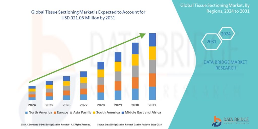
Growth Rate of Tissue Sectioning Market
The size of the global tissue sectioning market was estimated at USD 609.34 million in 2023 and is expected to grow at a compound annual growth rate (CAGR) of 6.40% from 2024 to 2031, to reach USD 921.06 million.
Learn More: https://www.databridgemarketresearch.com/reports/global-tissue-sectioning-market
Conclusion
An important development in pathology and histology is automated tissue sectioning. By enhancing precision, efficiency, and consistency, these systems revolutionize the way tissue samples are prepared for analysis. The benefits of automated tissue sectioning extend to improved diagnostic accuracy, accelerated research, and enhanced patient care. As technology continues to evolve, the integration of automated systems in histology laboratories will undoubtedly play a pivotal role in advancing medical science and improving healthcare outcomes.